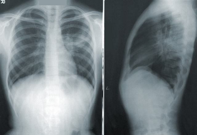
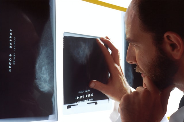
Chest X-ray is the most commonly used imaging test. Thanks to the use of X-rays (on film or in digital quality), the heart, lungs, respiratory tract, vessels and chest bones, as well as fragments of the spine are imaged in a non-invasive way. Performing an X-ray examination involves the body’s exposure to a small dose of ionizing radiation. Can cancer be seen on an x-ray?
Chest x-ray
Chest x-ray is an imaging study that involves short-term X-ray irradiation. Thanks to X-ray examination it is possible to visualize the airways, lungs, spine fragments, heart and chest bones. Irradiation creates an image of the chest and internal organs located in this area of the body.
When should a chest x-ray be performed?
Chest x-ray is most often performed when the following symptoms appear:
- shortness of breath,
- an alarming or persistent cough,
- chest pain or injury within it,
- chest injury,
- bleeding when you cough,
- esophageal bleeding.

X-ray examination is performed as a diagnostic examination in family medicine, cardiology and pulmonology. Chest x-ray is helpful in diagnosing and assessing the effectiveness of treatment:
- pneumonia,
- insufficiency and other heart diseases,
- emphysema,
- lung cancer
- interstitial changes,
- lung cancer (malignant and benign),
- cancer metastases from other organs,
- tuberculosis.
Lung cancer
This extremely dangerous disease is the most common malignancy in the world. Unfortunately, advanced changes are diagnosed in lung X-rays – it is practically impossible to visualize tumors smaller than 1 cm in diameter. Computed tomography is a much more sensitive test for lung cancer.
A change in the chest X-ray, suggesting a suspicion of cancer, as with pneumonia, is parenchymal shadowing. It is usually smaller and more “localized”, with clearer boundaries than inflammatory infiltrate. In order to make an accurate diagnosis, computed tomography, bronchoscopy and / or biopsy are necessary.


